2022
  • Yi-Teng Hsiao, Tsai-Ying Wu, Bo-Kuan Wu, Shi-Wei Chu, and Chia-Lung Hsieh*, Optics Express 30, 45233–45245 (2022) Website
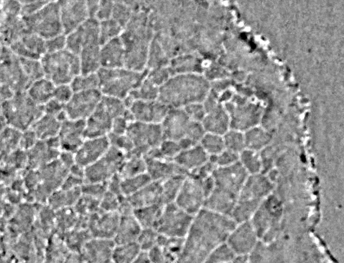
    Spinning disk interferometric scattering confocal microscopy captures millisecond timescale dynamics of living cells

 
Interferometric scattering (iSCAT) microscopy is a highly sensitive imaging technique that uses common-path interferometry to detect the linear scattering fields associated with samples. However, when measuring a complex sample, such as a biological cell, the superposition of the scattering signals from various sources, particularly those along the optical axis of the microscope objective, considerably complicates the data interpretation. Herein, we demonstrate high-speed, wide-field iSCAT microscopy in conjunction with confocal optical sectioning. Utilizing the multibeam scanning strategy of spinning disk confocal microscopy, our iSCAT confocal microscope acquires images at a rate of 1,000 frames per second (fps). The configurations of the spinning disk and the background correction procedures are described. The iSCAT confocal microscope is highly sensitive—individual 10 nm gold nanoparticles are successfully detected. Using high-speed iSCAT confocal imaging, we captured the rapid movements of single nanoparticles on the model membrane and single native vesicles in the living cells. Label-free iSCAT confocal imaging enables the detailed visualization of nanoscopic cell dynamics in their most native forms. This holds promise to unveil cell activities that are previously undescribed by fluorescence-based microscopy.

Link to the paper: https://doi.org/10.1364/OE.471935