
Advanced Microscopy
We develop advanced optical techniques that detect linear scattered light from small particles for studying important physical and chemical processes in living systems. The high sensitivity and the fast acquisition rate of our optical systems enable direct observation of the rapid motions of individual small particles in three dimensions.

- DETAIL -
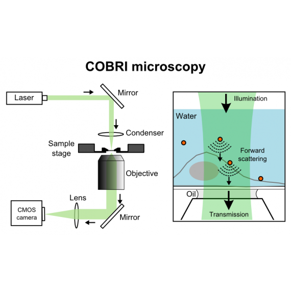
We believe the next-generation optical microscope technique should exploit the most basic light-matter interaction, that is, to take advantage of linear scattering and absorption (extinction) of light for image formation. In many circumstances, scattering-based approaches avoid the aforementioned limitations of the fluorescence method. A linear scattering signal is stable and indefinite, facilitating long-term observation. The strength of a linear scattering signal can be increased by raising illumination intensity. In addition, given a sufficient sensitivity, scattering-based imaging requires no label, allowing us to study living systems in their most native forms. To enhance the sensitivity of scattering-based imaging, we design the optical system and detect the signal by widefield interferometry (e.g., iSCAT microscopy and COBRI microscopy). Our current methods have the sensitivity to directly observe the dynamics of individual particles of 10 nm. In addition to the high sensitivity, the interferometric measurements also enable high precision at an ultrahigh image acquisition rate (100,000 frames per second). The ultrahigh speed and ultrahigh precision of scattering-based optical microscopy offers the opportunity to investigate dynamics in living systems with unprecedented clarity.
We believe that the forthcoming optical microscope technology should exploit the fundamental interaction between light and matter, specifically linear scattering and absorption, to create images. Scattering-based methods can avoid the limitations of fluorescence techniques in many situations. Linear scattering signals are stable and indefinite, allowing for high-speed and long-term observation. Additionally, scattering-based imaging can be label-free, allowing for the study of living systems in their natural forms. To enhance the sensitivity of scattering-based imaging, we design the optical system and detect the signal by widefield interferometry (e.g., iSCAT microscopy and COBRI microscopy). Our current methods have the capability to directly observe the dynamics of individual particles as small as 10 nm, with an ultrahigh image acquisition rate of 100,000 frames per second. The ultrahigh speed and precision of scattering-based optical microscopy offer an unparalleled opportunity to study dynamics in living systems with exceptional clarity.
More information:
iSCAT microscopy at 500,000 Hz
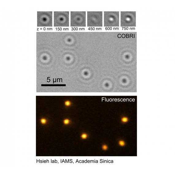
COBRI microscopy for single virus tracking at 100,000 Hz
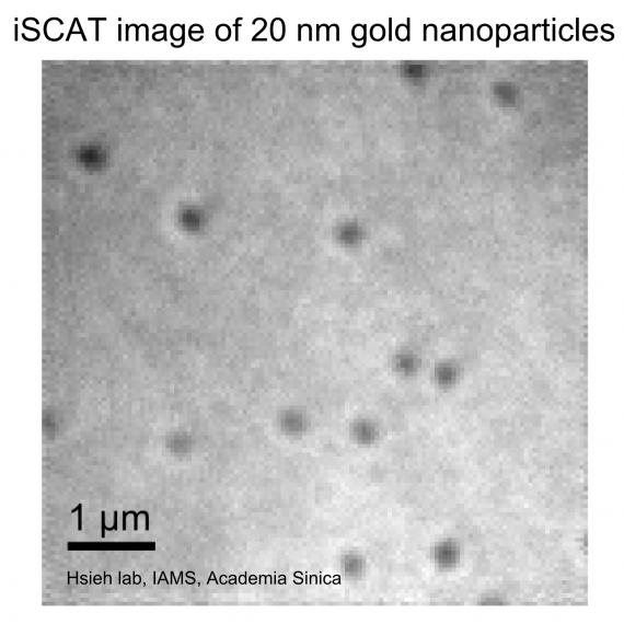
High-speed imaging and tracking of 10 nm gold nanoparticles
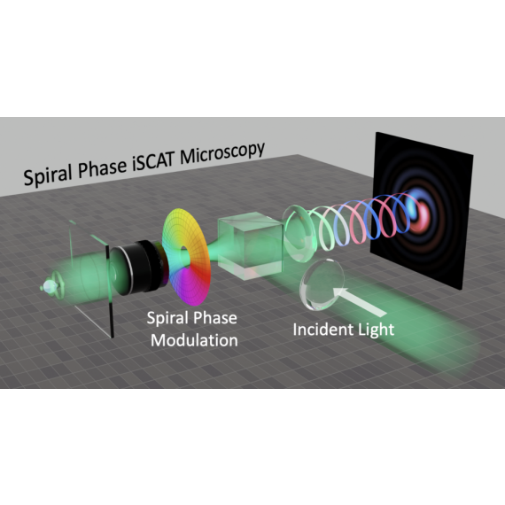
Spiral-phase iSCAT: iPSF engineering + iSCAT