
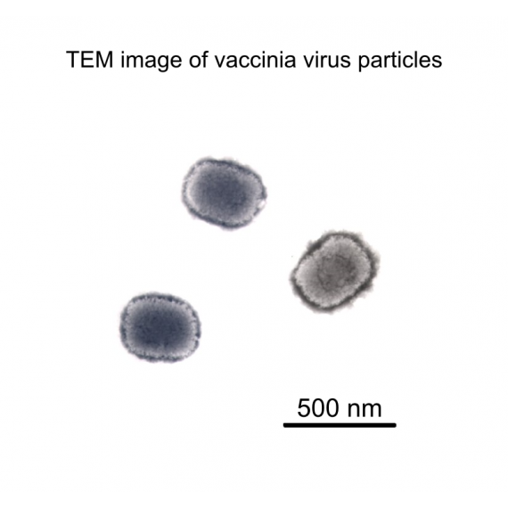
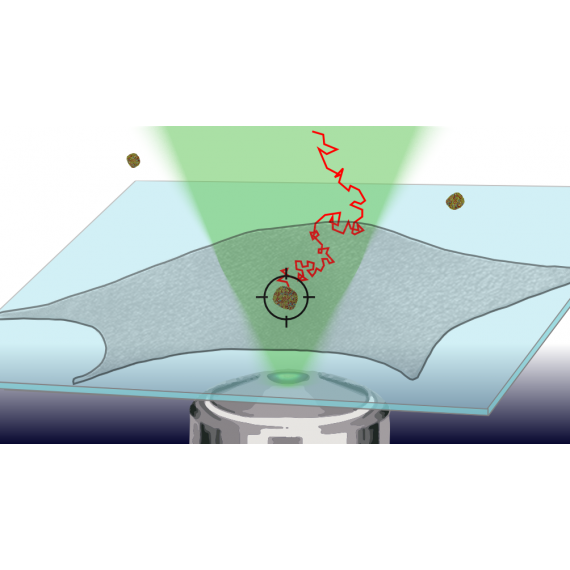
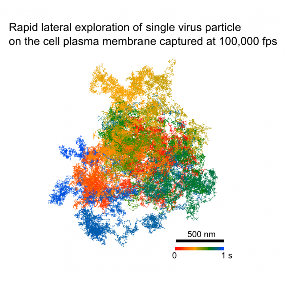
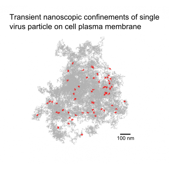
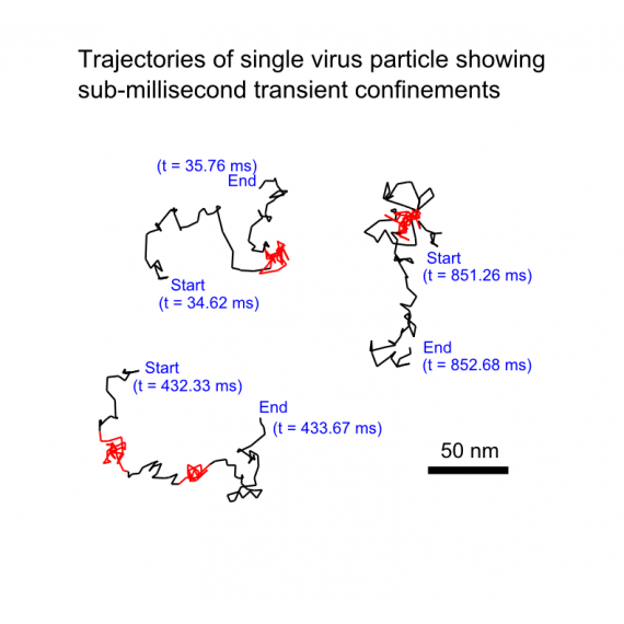
To address these questions, our lab and Dr. Wen Chang's lab at the Institute of Molecular Biology, Academia Sinica have been studying virus-receptor interaction in the early stages of viral infection using a combination of molecular biology approaches and advanced optical microscopy. We have used an ultrahigh-speed optical technique developed in our lab to capture the dynamic process of single vaccinia virus particles attaching to the cell plasma membrane with unprecedented spatial precision and temporal resolution (100,000 frames per second). By observing the virus-membrane interaction at the molecular length scale and microsecond timescale for the first time, we discovered that the vaccinia virus particle is initially confined within a zone of hundreds of nanometers in diameter and laterally explores the plasma membrane at a very high diffusion coefficient while transiently getting trapped at numerous nanoscopic sites within the confined zone. These sub-millisecond nano-confinements might be the result of virus-receptor interaction.
More about single virus tracking on cell plasma membrane: "Coherent Brightfield Microscopy Provides the Spatiotemporal Resolution To Study Early Stage Viral Infection in Live Cells", ACS Nano (2017).
Virus and Membrane
Disease outbreaks caused by viral infections have become a significant threat to our well-being. To combat such diseases, specially engineered virus particles are utilized as vaccines. Therefore, it is essential to comprehend the mechanisms through which viruses infect cells and initiate immune responses. We employ advanced optical microscope techniques to reveal virus-membrane interactions with ultra-high spatiotemporal resolution, enabling us to better understand and combat disease outbreaks caused by viral infections.
- DETAIL -